CRYOSCAN 2200
multifunctional scanning probe microscope
New possibilities for the study of surface morphology and local properties with subnanometer space resolution
Scanning probe microscopy in the 1,8 Ê- 350 K temperature range
- atomic force microscopy
- scanning tunneling microscopy
- scanning resistance microscopy
- electrostatic microscopy
- magnetic force microscopy
- nano lithography
- remote control via Internet

Technical parameters
| Cryogenic system |
| Temperature interval , K |
1,8 - 350 |
| Sample space diameter , mm |
40 |
| Helium reservoir , l |
2,2 or 4,2 |
| Nitrogen reservoir , l |
2,5 or 3,5 |
| Temperature stability in the interval 4 - 40, K* |
±0,04 |
| Temperature stability in the interval 40 - 300, K |
±0,2 |
| Cooling down time (to 4,2 K), min |
30 |
| Helium consumption for 4.2 K cooling down , l |
1,4 |
| Sample change time , min |
5 |
| Helium consumption at 4,2 K, l/hour |
<0,1 |
| Operation time at 1.8 Ê |
About 6 hours |
| Cryostat dimensions |
Diameter 200 mm, 750 mm height |
| Weight
, kg |
12 |
Probe microscopy
System for precise scanning:
Scanner FSS-3:
scanning range — up to 3 ?
step discrete — 0,01 nm
scanning rate max. — 30 Hz
typical thermal drift — less than 0,1 nm/sec
Scanner FSS-15:
scanning range — up to 15 ?
step discrete — 0,03 nm
scanning rate max. — 30 Hz
typical thermal drift — less than 1 nm/sec
sample orientation — horizontal
sample size — up to 10 mm in diameter and 5 mm in height
probe -sample approach — truly vertical (no lateral shift)
initial probe-sample approach - 4 mm by step motor
probe vertical displacement discrete during initial approach — 30 nm
Scanning tunneling microscopy mechanical head
STM modes: constant height, constant mode, spectroscopy, nanolithography
I(U) and I(Z) dependencies in selected regions
Resolution – atomic on graphite
Tunneling current — 50 pA-50 nA
Bias voltage range — ±10 V
Atomic force microscopy head
AFM modes: constant force, constant current, friction
Resolution — atomic on mica
|
Measurements in resonant modes
Nanolithography
Magnetic force microscopy mechanical head
Interleave and lift modes for contact and resonance modes
Force sensitivity – up to 10-11 N
Single magnetic domains space resolution
Cantilevers with different magnetic coatings
Simultaneous acquisition of topography and magnetic profile data
Electronic control unit
12 DAC (16 bit, 10 ?sec), 2 8-channels ADC (16 bit, 10 ?sec). DSP digital feedback. Embedded direct frequency synthesizer (0,01 Hz – 10 MHz)
Software
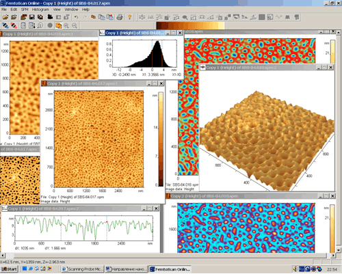
Microscope control software (master software) and data analysis software (client software) for Windows XP.
Master software – local or remote control (network, Internet).
Client software – local or remote real-time data acquisition, variety of filters, mathematical functions and algorithms for data analysis and 3D imaging.
Single Master and Multiple Clients operation. Embedded chat of intercommunication.
Detailed software description is placed at www.nanoscopy.net.
|
|


