Main Page
News
FAQ
Scan gallery
Links
Conditions of use
Privacy Statement
\.
Biology
Konkurs
Photo Contest
ScanOnline
Screeshot
Solid matter
Crystal growth
Lithography
Magnetic Imaging
Microdevices
Polymers
Quantum dots
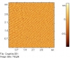
Graphite
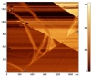
Domain boundary
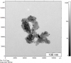
Red_silver_holesteric_np.4
Copy 1 of 01_1DEC
Copy 1 of 01_1DEC
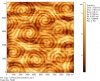
Dislocation network and Moire pattern
Dislocation network and Moire pattern: large scale
Pt-C2.jpg
graphite el st
2 um 89 nm Ar.002
Unbenannt.bmp
© 2018 Advanced Technologies Center